Right Ventricle 2 Chamber View
- cardiacmrihub
- Dec 1, 2025
- 2 min read
The right ventricle 2 chamber view provides a focused perspective for evaluating right heart anatomy and function. This guide covers the essential planning steps and clinical applications for acquiring this specialized cardiac MRI view.
2 Chamber Planning


Right Ventricle 2 Chamber View Planning
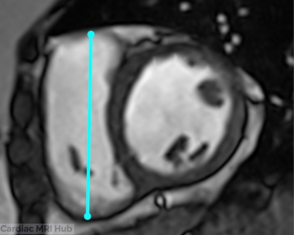
The 2 chamber view of the right ventricle is a vital imaging perspective in cardiac MRI, providing essential insights into the heart's anatomy and functionality. This view primarily focuses on the right atrium and right ventricle, allowing for a detailed assessment of their structure and any potential abnormalities.
Key Considerations for Planning
Orientation: The 2 chamber view is typically acquired from a plane that slices through the right atrium and right ventricle, ensuring clear visualization of both chambers.
Field of View: The field of view should adequately capture the right atrium and right ventricle, ensuring no critical structures are omitted.
Timing: Image acquisition should be synchronized with the cardiac cycle, preferably during diastole, to optimize visualization of the chamber sizes and function.
Contrast Agents: Consider the use of contrast agents to improve the delineation of myocardial tissue and enhance diagnostic precision.
Clinical Applications
Evaluation of right ventricular size and function.
Assessment of right atrial dimensions and morphology.
Identification of structural heart disease.
Monitoring of right ventricular hypertrophy or dilation over time.
Key Features of the 2 Chamber View
Right Atrium: Detailed analysis of right atrial size and any structural abnormalities.
Right Ventricle: Assessment of right ventricular volume, wall motion, and overall cardiac function.
Conclusion
Mastering the right ventricle 2 chamber view requires careful attention to patient positioning, plane orientation, and timing. By following these planning guidelines, technologists can consistently obtain high-quality images that support accurate diagnosis and treatment of right heart conditions.


Comments